
Educational scope: Teaching starting points, concepts and arithmetic for learning; not a ventilator simulator or a prescription for a real patient. Free, with no account or email needed.
Written by Dr. Ezekiel Aluda Osolika, emergency physician (MBChB, FEBEM). Clinical content reviewed by the author on 24 October 2026; review covered the case histories, answers, explanations, calculations, image interpretation, annotation guides and teaching diagrams. This is author review, not an independent second review. All patients and orders are fictional. Content version 2026-10-24-1. Spotted an error? Report a correction and name the case ID.
Start ventilator settings practice
Twelve fictional scenarios, one scored concept question each. Two answers open a small arithmetic sandbox; none of them predicts a real patient’s blood gas or saturation.
The interactive practice needs JavaScript. Every case, answer and explanation is also written out in the worked answers below.
How to practise
- Separate oxygenation from ventilation. Inspired oxygen, effective alveolar ventilation, lung mechanics and the patient’s condition are related but not interchangeable.
- Use predicted rather than measured body weight when discussing the named lung-protective reference approach. A calculation provides a teaching reference, not a complete ventilator prescription.
- Read pressure and flow patterns in context. Valid plateau assessment, patient effort, expiratory time and total PEEP matter when interpreting the measurements.
- When an alarm accompanies deterioration, assess the patient and obtain appropriate help. Do not make the alarm disappear by disabling protection or adjusting a limit without addressing the problem.
- The diagrams are explanatory schematics, not patient recordings or a validated physiologic simulator. Clinical ventilator adjustments require the trained treating team and local protocol.
For nurses and paramedics: use these cases for recognition, bedside checks, communication and escalation within your local competence. Nothing here extends your scope or replaces supervised ventilator training.
Predicted body weight uses the ARDSNet reference equation in its exact inch form: PBW (male) = 50 + 2.3 × (height in inches − 60). The commonly rounded metric version gives a nearby result; case 3 uses the inch form throughout.
Initial settings: mode, tidal volume, rate, PEEP and FiO2
- Mode. Volume-controlled assist-control is a common first mode because it guarantees each breath’s volume; pressure control is a reasonable alternative where it is the local norm. Pressure support and SIMV-type modes are generally for later, when the patient is breathing usefully.
- Tidal volume. 6–8 mL/kg predicted body weight, at the lower end when the lungs are injured or at risk.
- Rate. Often 14–18 breaths/min. A patient who was compensating for a severe metabolic acidosis before intubation needs a minute ventilation close to their own; a low default rate can drop the pH quickly.
- PEEP. 5 cmH2O is the usual starting point, increased for hypoxaemia with recruitable lung.
- FiO2. Start high immediately after intubation, then titrate down to the SpO2 target, commonly 94–98%, or 88–92% for patients at risk of hypercapnic respiratory failure.
- Then check. Measure a plateau pressure, look at the flow waveform for complete exhalation, confirm end-tidal CO2, and take a blood gas after about 20–30 minutes.
These are common teaching starting points for an adult after intubation, not a prescription. Your local protocol, the patient’s condition and the senior or remote adviser decide the actual settings.
What tidal volume should I start at?
Start at 6–8 mL/kg of predicted body weight, which is calculated from height and sex, not from measured weight. For a man 175 cm tall, predicted body weight is about 71 kg, so 6–8 mL/kg is roughly 420–565 mL. For a woman 160 cm tall it is about 52 kg, or roughly 315–420 mL. Use the lower end in lung injury, then confirm the plateau pressure stays at or below 30 cmH2O. Case 3 works through the calculation.
Lung-protective ventilation and why it matters
Large breaths overstretch the lung that is still open and add injury to the disease that caused the intubation. In the ARDS Network trial, ventilating with 6 rather than 12 mL/kg predicted body weight reduced mortality. The working principles that follow from it are:
- size tidal volume to predicted body weight;
- keep the plateau pressure at or below 30 cmH2O and keep the driving pressure (plateau minus PEEP) as low as practical;
- accept a modest rise in PaCO2 when the pH remains acceptable, rather than chasing a normal CO2 with large breaths;
- use enough PEEP and FiO2 to meet the oxygen target, following a table or local protocol.
Oxygenation or ventilation: which dial do you turn?
Two separate problems have two separate sets of controls.
- Low oxygen: the oxygenation dials are FiO2 and PEEP, which raise inspired oxygen and mean airway pressure and keep alveoli open.
- High CO2: the ventilation dials are rate and tidal volume, which together set minute ventilation. Raise the rate before the tidal volume if the plateau pressure is near its limit, and watch expiratory time in asthma and COPD, where a faster rate can trap gas.
Before turning any dial, check that the problem is not the tube, the circuit or the patient. Case 8 and case 9 practise the distinction.
Troubleshooting the high-pressure alarm
- Look at the patient first. If they are deteriorating, disconnect from the ventilator and hand-ventilate with 100% oxygen. If bagging is easy, suspect the ventilator or circuit; if bagging is hard, the problem is the tube or the patient.
- Work through DOPES: Displacement of the tube, Obstruction (secretions, kinking, biting), Pneumothorax, Equipment failure, and Stacked breaths (gas trapping).
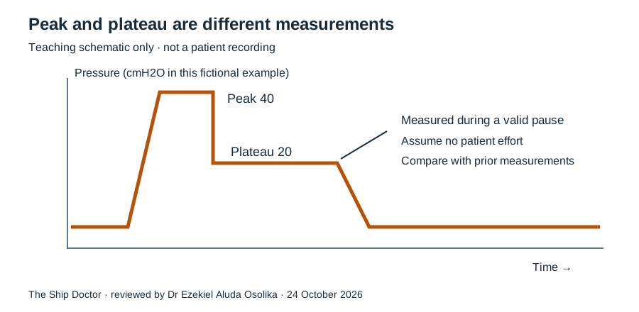
- Compare peak and plateau pressures. A high peak with a normal plateau points to airway resistance: a kinked or blocked tube, secretions or bronchospasm. A high peak and a high plateau points to stiff lungs or chest: pneumothorax, endobronchial intubation, pulmonary oedema, abdominal distension or gas trapping.
- Never silence the alarm or raise the limit to make it stop before the cause is found.
Teaching waveforms
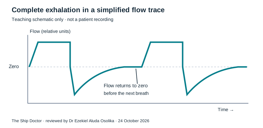
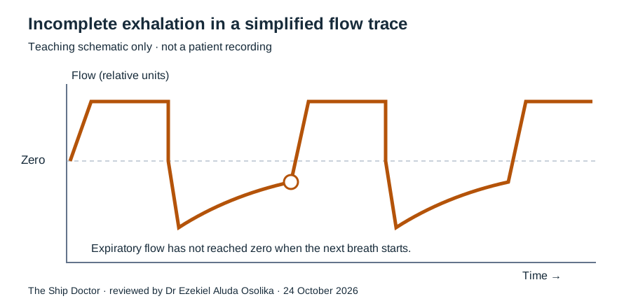
Three simplified schematics support cases 4 and 6. They are drawn to explain a concept, are not patient recordings, and are not drawn to an exact time scale. Read them with the stated assumptions: a passive patient, a valid inspiratory pause and no patient effort.
Practice scenarios with worked answers
Every case in full, with the correct answers and the reasoning. Open one when you have committed to your own answer.
Foundations: cases 1–3
Case 1 Minute ventilation arithmetic Foundations
A fictional controlled ventilation example has tidal volume 450 mL and respiratory rate 16/min. All delivered breaths have the stated volume.
Question 1: Which answer best fits this teaching scenario?
Answer: Minute ventilation is 7.2 L/min
Worked answer
- Minute ventilation = tidal volume × rate.
- 450 mL × 16/min = 7200 mL/min = 7.2 L/min.
- This is total minute ventilation, not alveolar ventilation; it does not by itself predict PaCO2.
Arithmetic sandbox: minute ventilation
Minute ventilation = tidal volume × rate ÷ 1000 = L/min
Fictional arithmetic only. It shows what the numbers multiply to, not a recommended setting or a predicted blood gas.
Case 2 Dead space changes the meaning Foundations
Two fictional examples both deliver 6 L/min: A is 500 mL × 12/min and B is 300 mL × 20/min. Assume a fixed dead space of 150 mL per breath solely for this exercise.
Question 1: Which answer best fits this teaching scenario?
Answer: A has greater calculated alveolar ventilation: 4.2 versus 3.0 L/min
Worked answer
- Use (tidal volume − assumed dead space) × rate.
- A: (500 − 150) × 12 = 4200 mL/min. B: (300 − 150) × 20 = 3000 mL/min.
- Dead space is not fixed in real disease. This simplified comparison does not establish the correct setting or predict an actual blood gas.
Case 3 Predicted body weight is not measured weight Foundations
For a fictional adult using the ARDSNet male reference equation, height is 180 cm. Calculate predicted body weight and a 6 mL/kg teaching tidal volume. Actual body weight is 110 kg.
Question 1: Which answer best fits this teaching scenario?
Answer: Predicted body weight is about 75.0 kg and 6 mL/kg is about 450 mL
Worked answer
- The exact inch equation is PBW = 50 + 2.3 × (height in inches − 60).
- 180 cm ÷ 2.54 = 70.866 inches; PBW ≈ 74.99 kg.
- 6 × 74.99 ≈ 450 mL. The commonly rounded metric coefficient gives a nearby result; use one convention consistently.
- A calculated reference volume still requires clinical individualisation and pressure assessment.
Pressures and waveforms: cases 4–6
Case 4 Peak rises but plateau is unchanged Advanced
In a fictional passive patient with constant inspiratory flow, peak pressure rises from 25 to 40 cmH2O while plateau remains 20. Measurements are valid and total PEEP is unchanged.
Question 1: Which answer best fits this teaching scenario?
Answer: An increase in airway resistance is a leading explanation
Worked answer
- The larger peak to plateau difference suggests increased resistive pressure under the stated conditions.
- Assess the patient, airway and circuit, including possible kinking, secretions or bronchospasm.
- The pattern guides assessment; it does not independently diagnose the cause. Plateau interpretation requires an appropriate pause without confounding effort.
Case 5 Both pressures rise Advanced
In a fictional passive patient, peak rises from 25 to 35 and plateau rises from 20 to 30 cmH2O. Delivered volume, total PEEP and inspiratory flow are unchanged.
Question 1: Which answer best fits this teaching scenario?
Answer: A change in respiratory system compliance is a leading explanation
Worked answer
- Under these controlled assumptions, a higher plateau at the same volume and total PEEP suggests reduced respiratory system compliance.
- Assess patient and circuit, and consider lung and chest wall factors rather than jumping to one diagnosis.
- Use a valid plateau measurement and evaluate the whole clinical picture.
Case 6 Expiration has not finished Alarms
An expiratory flow schematic remains below the zero flow baseline when the next breath starts.
Question 1: Which answer best fits this teaching scenario?
Answer: Incomplete exhalation suggests air trapping and possible intrinsic PEEP
Worked answer
- Expiratory flow returning to zero suggests that flow has ceased before the next inspiration.
- Failure to reach zero may indicate incomplete exhalation and air trapping; assess the patient and measurement context.
- Management requires clinician assessment of expiratory time, obstruction and the ventilatory strategy. Increasing the rate can make expiratory time shorter.
Alarms and gas exchange: cases 7–8
Case 7 A low pressure alarm Alarms
A fictional ventilated patient suddenly triggers a low pressure alarm and has a marked fall in measured exhaled volume.
Question 1: Which answer best fits this teaching scenario?
Answer: Assess the patient immediately and check for disconnection or a significant leak while obtaining help
Worked answer
- A disconnection or leak is one possible explanation for loss of pressure and exhaled volume.
- Look at the patient first, call for appropriate help and assess the airway and breathing circuit.
- If ventilation is failing, a trained clinician must provide appropriate alternative support according to the emergency protocol.
Case 8 Oxygenation and CO2 clearance Foundations
A learner says that raising FiO2 should correct a high PaCO2 in every ventilated patient.
Question 1: Which answer best fits this teaching scenario?
Answer: FiO2 primarily changes inspired oxygen; CO2 clearance depends on effective alveolar ventilation and physiology
Worked answer
- FiO2 addresses inspired oxygen concentration.
- CO2 clearance depends on effective alveolar ventilation and factors including dead space and CO2 production.
- Oxygenation and ventilation need separate assessment. Avoid assigning a one size fits all response to a single control change.
Advanced: cases 9–12
Case 9 Why increasing the rate is not always harmless Advanced
A fictional obstructive lung disease scenario shows incomplete exhalation. A learner proposes increasing respiratory rate because the PaCO2 is high.
Question 1: Which answer best fits this teaching scenario?
Answer: A higher rate can shorten expiratory time and worsen air trapping; reassess the complete strategy
Worked answer
- Higher rate may increase total minute ventilation but shorten the time available for expiration.
- In obstructed lungs, the change can worsen dynamic hyperinflation.
- The appropriate strategy depends on the patient and must be directed by a trained clinician.
Arithmetic sandbox: time per breath
Total cycle time = 60 ÷ rate = seconds per breath
The whole cycle shrinks as the rate rises, so less time is left for exhalation unless inspiratory time also changes. The rate does not set inspiratory time automatically, and this sandbox does not predict a PaCO2.
Case 10 PEEP can have more than one effect Advanced
In a fictional scenario, PEEP is increased. Oxygenation appears better, but blood pressure falls.
Question 1: Which answer best fits this teaching scenario?
Answer: Reassess both respiratory and haemodynamic effects; improved saturation is not the only outcome
Worked answer
- PEEP can affect recruitment, intrathoracic pressure and haemodynamics.
- A change in blood pressure needs prompt assessment rather than automatic attribution to one cause.
- The team should review the patient, ventilator measurements and response together.
Case 11 A high pressure alarm is a patient assessment task Alarms
A fictional ventilated patient has a high pressure alarm, falling saturation and agitation.
Question 1: Which answer best fits this teaching scenario?
Answer: Call for help and assess the patient, airway and circuit immediately
Worked answer
- The combination of an alarm and deterioration is urgent.
- Possible contributors include obstruction, circuit problems, dyssynchrony and changing lung mechanics; assess rather than assume.
- If support is ineffective, trained clinicians follow the service’s emergency ventilation approach.
Case 12 Readiness is more than one normal gas Advanced
A fictional patient has an improved blood gas and low oxygen requirements. The learner wants to remove ventilatory support based only on those numbers.
Question 1: Which answer best fits this teaching scenario?
Answer: Readiness also requires clinical assessment, an appropriate liberation process and airway protection evaluation
Worked answer
- Gas exchange is one part of a broader assessment.
- A supervised liberation process also considers the underlying illness, respiratory effort, haemodynamics and ability to protect the airway.
- Follow the local trained team’s pathway; this practice lab cannot decide readiness for an individual.
Ventilating a patient you cannot transfer
At sea, offshore or at a remote site, a ventilated patient may stay with you for hours or days. The settings matter, and so does everything around them:
- Oxygen is finite. Titrate FiO2 to the target rather than leaving it high, and know your supply. A gas-driven transport ventilator may also use oxygen to drive itself. The oxygen burn-rate calculator turns flow into hours remaining.
- Monitor continuously. Waveform capnography is the earliest warning of a displaced or blocked tube. Recheck tube position after every move.
- Prevent harm over time. Keep the head of the bed up about 30 degrees, check cuff pressure, humidify the circuit, suction as needed, sedate to a target and protect pressure areas.
- Escalate with numbers. Give the remote adviser the mode, set and measured values, plateau pressure, SpO2, end-tidal CO2 and the latest gas, not just “the patient is ventilated”.
The 72-hour maritime ICU covers sustaining a ventilated patient far from definitive care.
Questions about this practice
Does changing a control predict a real patient’s blood gas?
No. The small sandboxes show stated arithmetic and qualitative tradeoffs, not validated physiologic predictions.
Can nurses and paramedics use this?
Yes, for concepts, recognition, communication and role-appropriate practice. The page does not extend anyone’s clinical scope.
Where should I start?
Begin with minute ventilation and dead space, then try the alarm cases. Review ABG fundamentals alongside the more advanced material.
Educational disclaimer
Educational use only. These ventilation scenarios and schematics teach concepts only. No output on this page is a ventilator prescription. Actual changes require trained supervision and your local protocol. Educational fictional practice only. Not patient-specific advice or an assessment of clinical competence. Scores are informal feedback on one practice attempt; no pass mark, certificate or competence decision is made from them. Follow your supervision, scope of practice and local protocols.
Sources and further reading
- AARC — Clinical practice guideline patient ventilator assessment 2024
- ARDS Network — Lower tidal volume trial 2000
- ARDS Network protocol card hosted by George Washington University
- AARC — Spontaneous breathing trials for liberation from adult mechanical ventilation 2024
Reference ranges, formulae and pathways vary between laboratories, services and countries. Use your own local guidance in practice.